


 59 / 127

57
Capítulo 12. O que é um buraco lamelar?
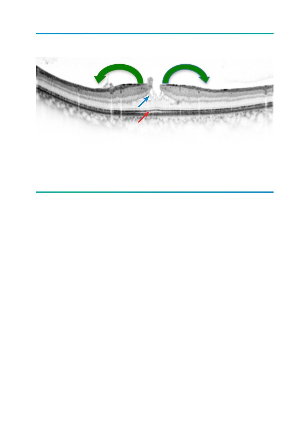
Figura 2.
Imagem de SD-OCT de buraco lamelar onde se pode observar a típica separação horizontal intra-retiniana,
entre a camada plexiforme externa e camada nuclear externa (seta azul) e a manutenção da integridade da
camada dos fotorreceptores (seta vermelha), importante para o diagnóstico diferencial com pseudoburaco
macular. Repare na tracção centrífuga exercida pela MER (seta verde).
REFERÊNCIAS
1.
Gass JD. Lamellar macular hole: a complication
of cystoid macular edema after cataract extrac-
tion: a clinicopathologic case report. Trans Am
Ophthalmol Soc 1975;73:231-50.
2.
Duker JS, Kaiser PK, Binder S, de Smet MD,
Gaudric A, Reichel E. The international vitreo-
macular traction study group classification of
vitreomacular adhesion, traction, and macular
hole. Ophthalmology 2013;120(12):2611-9.
3.
Witkin AJ, Ko TH, Fujumoto JG, Schuman JS,
Baumal CR, Rogers AH. Redefining lamellar
holes and the vitreomacular interface: an ul-
trahigh-resolution optical coherence tomogra-
phy study. Ophthalmology 2006:113:388-97.
4.
Xirou T, Kidess A, Kourentis C, Xirou V, Feretis
E, Kabanarou SA. Lamellar macular hole for-
mation following vitrectomy for rhegmatoge-
nous retinal detachment repair. Clin Ophthalmol
2012;6:571-4.
5.
Takahashi H, Kishi S. Tomographic features of
a lamellar macular hole formation and a lamellar
hole that progressed to a full-thickness macular
hole. Am J Ophthalmol 2000;130:677-9.
6.
Haouchine B, Massin P, Tadayoni R, Erginay
A, Gaudric A. Diagnosis of macular pseu-
doholes and lamellar macular holes by opti-
cal coherence tomography. Am J Ophthalmol
2004:138:732-9.
7.
Figueroa MS, Noval S, Contreras I. Macular
structure on optical coherence tomography
after lamelar macular hole surgery and its cor-
relation with visual outcome. Can J Ophthalmol
2011;46:491-7.
8.
Sebag J, Wang MY, Nguyen D, Sadun AA.
Vitreopapillary adhesion in macular diseases.
Trans Am Ophthalmol Soc 2009;107:35-44.
9.
Chen JC, Lee LR. Clinical spectrum of lame-
lar macular defects including pseudoholes and
pseudocysts defined by optical coherence to-
mography. Br J Ophthalmol 2008;115:884-6.
10.
Meyer CH, Rodrigues BE, Mennel S, Sch-
midt JC, Kroll P. Spontaneous separation
of epireti- nal membrane in young subjects:
personal observations and review of the li-
terature. Graefe’s Arch Clin Exp Ophthalmol
2004:242:977-85.