


 94 / 127

92
A interface vítreo-retiniana
AVM, TVM e BM
25 Perguntas e respostas
REFERÊNCIAS
1.
Imamura Y, Zweifel SA, Fujiwara T, Freund KB,
Spaide RF. High-Resolution Optical Coherence
tomography findings in optic pit maculopathy.
Retina 2010;30:1104-12.
2.
Gandorfer A, Kampik A. Role of vitreoretinal in-
terface in the pathogenesis and therapy of mac-
ular disease associated with optic pits. Ophthal-
mologe 2000;97:276-9.
3.
Hirakata A, Hida T, Wakabayashi T, Fukuda M.
Unusual Posterior Hyaloid Strand in a Young
Child with Optic Pit Maculopathy: Intraoperative
and Histopathological Findings. Jpn J Ophthal-
mol 2005;49(3):264-6.
4.
Christoforidis JB, Terrel W, Davidorf FH. Histo-
pathology of optic nerve pit-associated macu-
lopathy. Clin Ophthalmol 2012;6:1169-1174.
5.
Bonnet M. Serous macular detachment associ-
ated with optic nerve pits. Graefes Arch Clin Exp
Ophthalmol 1991;229(6):526-32.
6.
Montenego M, Bonnet M. Optic nerve pits:
clinical and therapeutic review of 21 cases. J Fr
Ophthalmol 1989;12(6-7):411-9.
7.
Postel EA, Pulido JS, McNamara JA, Johnson
MW. The etiology and treatment of macular de-
tachment associated with optic nerve pits and
related anomalies. Trans Am Ophthalmol Soc
1998;96:73-93.
8.
Taiel-Sartral M, Mimoun G, Glacet-Bernard A, De-
layre T, Coscas G. [Vitrectomy-laser-gas for treat-
ing optic disk pits complicated by serous macular
detachment]. J Fr Ophthalmol 1996;19(10):603-9.
9.
Mohammed OA, Pai A. Inverted autologous
internal limiting membrane for management of
optic disc pit with macular detachment. MEAJO
2013;20(4):357-9.
10.
Jalil A, Stavrakas P, Dhawahir-Scala FE, Patton
N. Drainage of subretinal fluid in optic disc pit
maculopathy using subretinal 42-gauge can-
nula: a new surgical approach. Graefes Arch
Clin Exp Ophthalmol 2010;248(5):751-3.
11.
Shukla D, Kalliath J, Tandon M, Vijayakumar
B. Vitrectomy for optic disk pit with macular
schisis and outer retinal dehiscence. Retina
2012;32(7):1337-42.
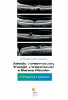
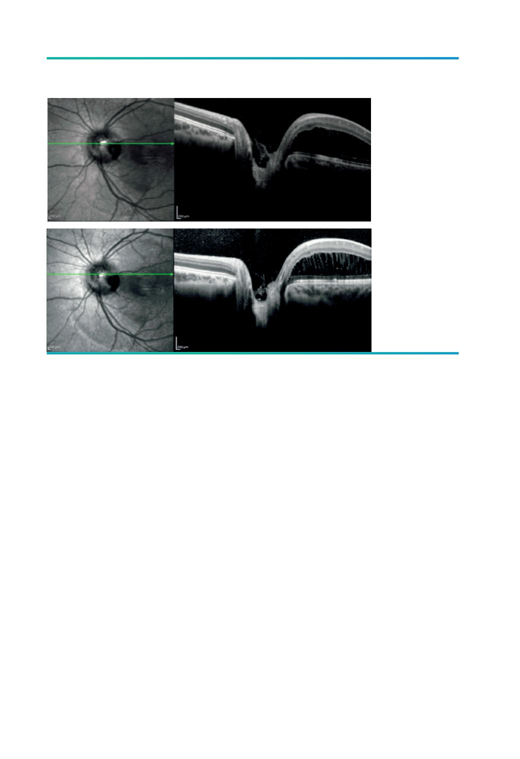
Figura 6.
OCT no bordo
superior da fosseta
colobomatosa do
paciente da figura 2
onde se evidencia
a presença de
adesão vítrea. O
aumento do ganho
na imagem inferior
permite identificar
melhor a adesão
vítrea, assim como
o estiramento
celular associado à
presença de fluido
na camada nuclear
externa.
A
B