


 110 / 127

108
A interface vítreo-retiniana
AVM, TVM e BM
25 Perguntas e respostas
REFERÊNCIAS
1.
Squirrel D, Ehrlich R. The use of Macular
Microperimetry in the assessment and
diagnosis of Macular Diseases. Retinal
Physician 2012;9(65):53-7.
2.
Seiple W, Rosen RB, Castro-Lima V, Garcia
PM. The Physics and Psycophysics of
Microperimetry. OVS 2012;89(8):1182-91.
3.
Chen, WC, Wang Y, Li XX. Morphologic and
functional evaluation before and after successful
macular hole surgery using spectral-domain
optical coherence tomography combined with
microperimetry. Retina 2012;32:1733-42.
4.
Parravano M, Oddone F, Boccassini B,
Chiaravalloti A, Scarinci F, Sciamanna M et
al. Functional and structural assessment of
lamellar macular holes. Br J Ophthalmology
2013;97:291-6.
5.
Chen JC, Lee LR. Clinical spectrum of lamellar
macular defcts including pseudoholes and
pseudocysts defined by optical coherence
tomography. Br J Ophthalmol 2008;92:1342-
6.
6.
Grimbert P, Lebreton O, Weber M. En
face optical coherence tomography and
microperimetry after internal limiting
membrane peeling for epiretinal membrane.
J Fr Ophtalmol 2014;37(6):434-41.
7.
Mayer WJ, Vogel M, Neubauer A, Kernt
M, Kampik A, Wolf A et al. Pars plana
vitrectomy and internal limiting membrane
peeling
in
epimacular
membranes:
correlation of function and morphology
across the macula. Ophthalmologica
2013;230(1):9-17.
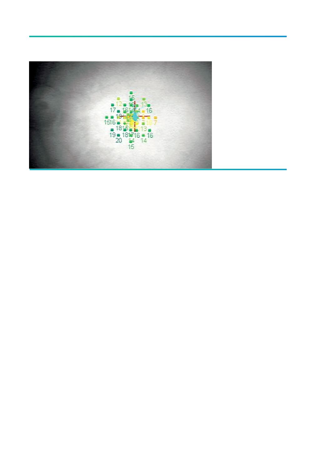
(Imagens cedidas pelo Dr. David Martins, Hospital de Setúbal, de doente operado em fevereiro de
2014. As microperimetrias foram efectuadas no Instituto Gama Pinto pela Dra. Luisa Colaço).
Figura 6.
Microperimetria MP1
pós-operatória com
sensibilidade média de
13.2 dB.